Intracellular Delivery Image Gallery
 |
 |
 |
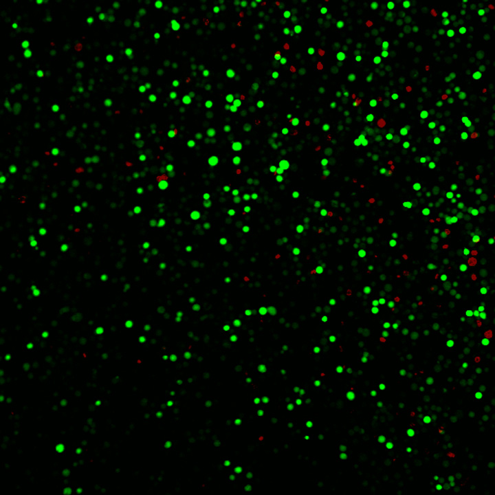
| A field of human prostate cancer cells after exposure to laser-activated carbon nanoparticles. The many green cells have taken up a model therapeutic compound, calcein, while the few red-stained cells are dead. Each of the green or red spots is a single cell. (Credit: Prerona Chakravarty, Georgia Tech) |
A field of human prostate cancer cells is shown after exposure to laser-activated carbon nanoparticles. The cell membranes have been stained red to assist in visualization. Each of the red circles is a single cell. (Credit: Prerona Chakravarty, Georgia Tech)
|
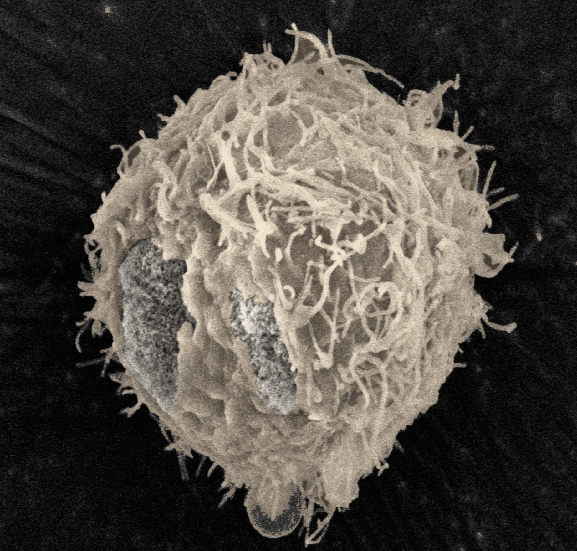
Scanning electron micrograph showing a prostate cancer cell immediately after exposure to ultrasound. Image has been color enhanced to show to the spot where the cell membrane has been removed. (Credit: Robyn Schlicher, Georgia Tech) |
 |
||
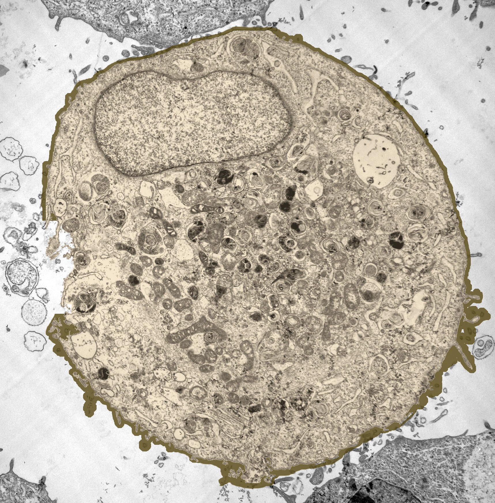
| Transmission electron micrograph showing a prostate cancer cell immediately after exposure to ultrasound. Image has been color enhanced to show to the spot where the cell membrane has been removed. (Credit: Robyn Schlicher, Georgia Tech) |